Laboratory / facilities & equipment
Nonlinear Microscopy setup
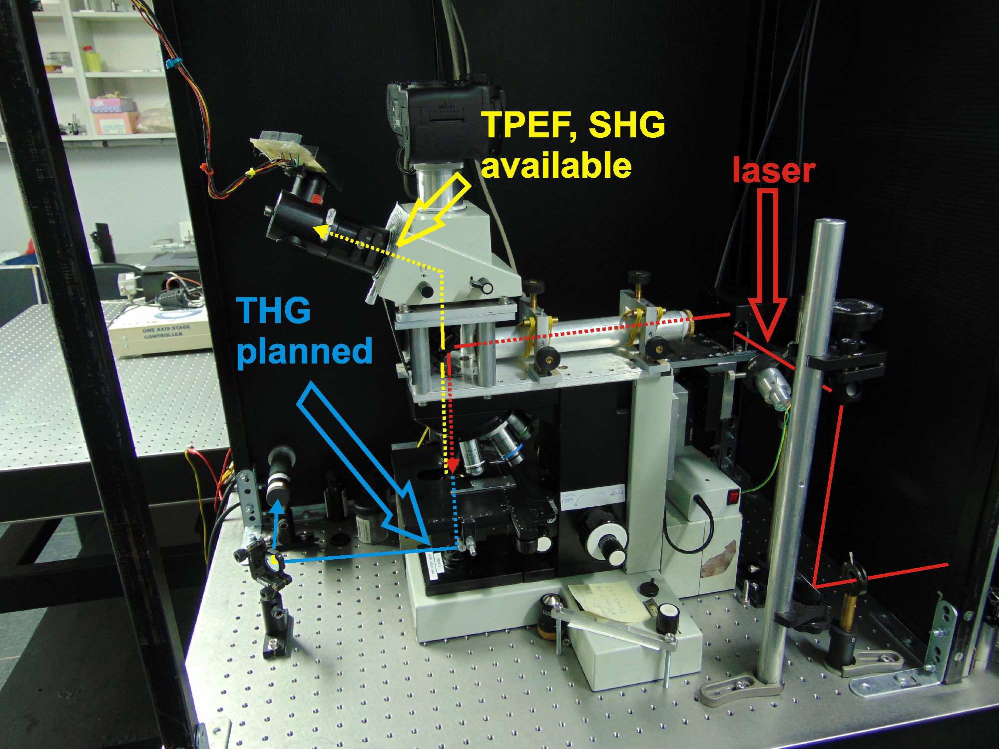
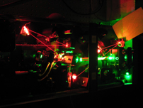
Our homemade experimental setup for nonlinear laser scanning microscopy (NLSM) have both, reflection and transmission detection arms, and consists of following imaging modalities:
- Two Photon Excitation Fluroescence - TPEF
-
excitation: 720-930nm and 1040nm
-
detection: selection of broad and narrow band filters in VIS region
- Second Harmonic Generation - SHG
-
polarization resolved
-
excitation: 840nm, 900nm, 1040nm
-
detection: 420nm, 450nm and 520nm
- Third Harmonic Generation - THG
-
excitation: 1040nm
-
detection: 347nm
In addition, there is possibility for sample processing: cutting, inscription of arbitrary patterns, selective photo bleaching.
More about the NLSM setup
Lasers
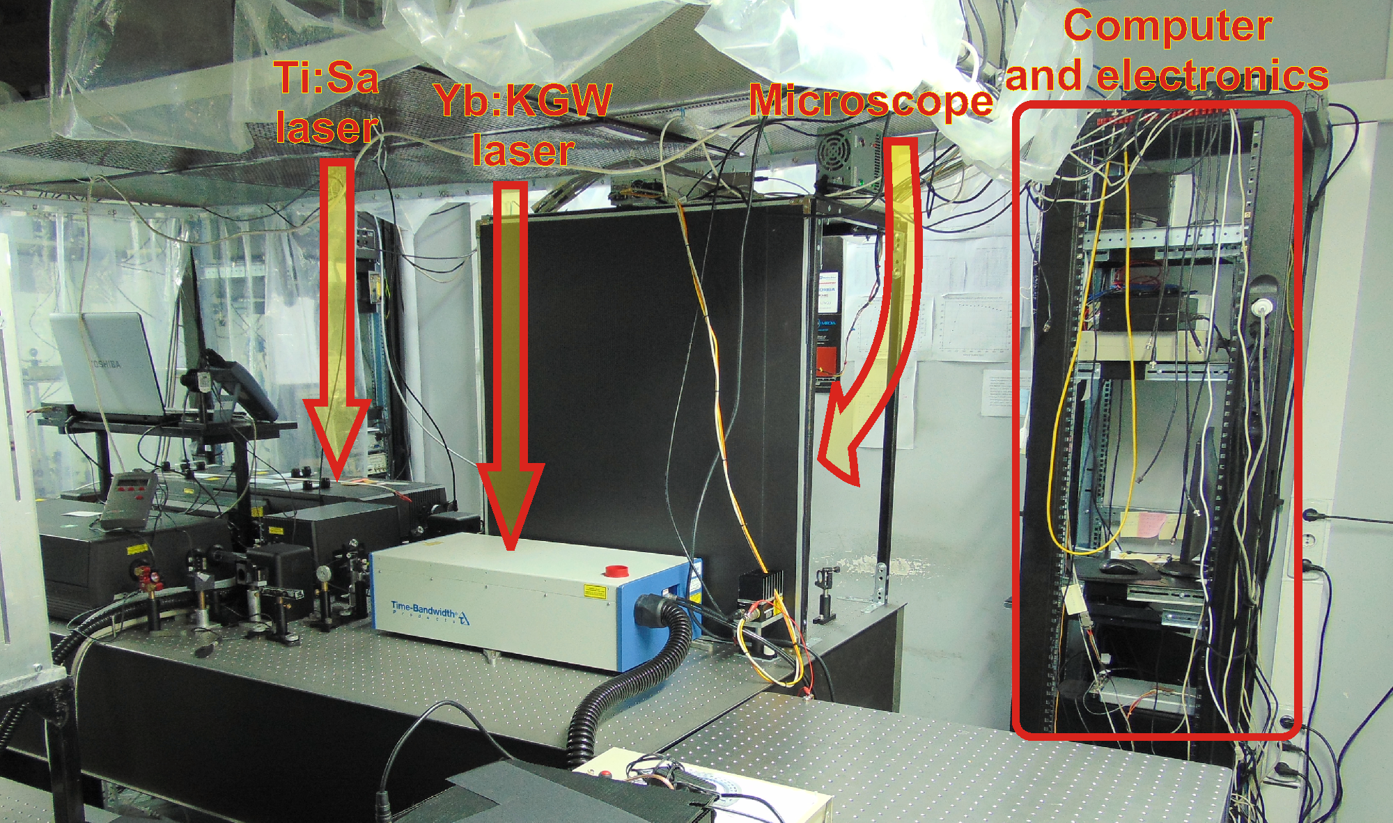
Laser sources used for excitation in NLSM:
-
Kerr lens mode locked Ti:Sa laser
- wavelength: 700-1000nm; pulse duration: 160 fs; repetition rate: 76 MHz; average power: 2 W
- Pulse picker for Ti:Sa laser
- repetition rate: single shot to 4 MHz
- Regenerative amplifier for Ti:Sa laser
-
pulse energy 6 µJ, repetition rate: 250 kHz; wavelength 800 nm; pulse duration: 200 fs
- SESAM mode locked Yb:KGW laser
- wavelength: 1040nm; pulse duration: 200fs; repetition rate: 83MHz; average power: 2W
More about the lasers
Optical autocorrelator
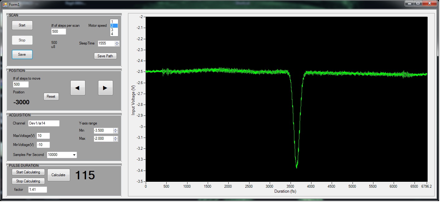
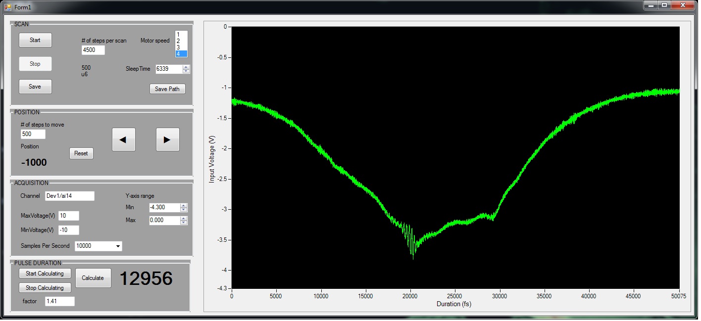
Our homemade optical autocorrelator enables measurements of ultrashort pulse durations in the range 10 fs - 100 ps.
More about the autocorrelator
Cooled CCD spectrometer
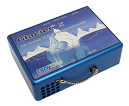
Portable, fiber coupled, USB spectrometer, coupled with NLSM setup, for very weak fluorescence signal measurements and spectral analysis upon the excitation in the arbitrary chosen point on the image.
More about the spectrometer
In progress
We are currently developing two more microscopic techniques:
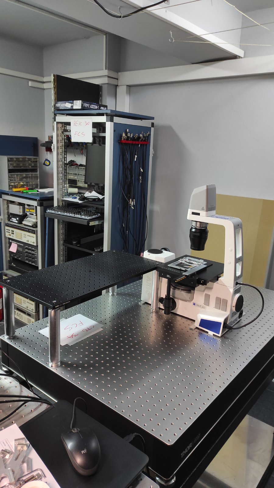
- Fluorescence correlation spectroscopy (FCS) - quantitative technique with single molecule sensitivity (through Hemmaginero project)
Further reading of FCS:
- Structured Illumination Microscopy (SIM) - imaging technique with super resolution (through Move For The Science - #PokreniNauku project)
Further readings on SIM:
 Nonlinear Microscopy setup
General. TPEF and SHG modalities are fully operational in back reflection arm, while the transmission arm for both THG and SHG is under development through the HEMMAGINERO project. Also, it is possible to perform polarization SHG imaging in backreflection with control of both, polarization of incident laser beam and polarization of SHG signal (analyzer).
Two Photon Excitation Fluroescence - TPEF is the most common effect/signal that is detected in NLSM. It is analogous to the
single photon excitation that is utilized in confocal and epifluroescent microscopy,
but in TPEF the fluorescent molecule is excited by absorbing two photons simultaneously.
Upon the two photon excitation, the fluorescence occurs in the same manner as in the single photon excitation.
NLSM set up with lasers
Objective lenses
| Type |
Magnification |
NA (immersion) |
Working distance |
Max field of view |
| Zeiss |
12.5 |
0.3 |
|
551 |
| Zeiss Plan-APOCHROMAT |
20 |
0.8 |
0.55 |
233.9 |
| Zeiss EC Plan-NEOFLUAR |
40 |
1.3 (oil) |
0.21 |
115.4 |
| Zeiss |
40 |
0.65 |
|
|
| Zeiss W Plan-Apochromat 2.5 |
40 |
1.0 (phys) |
2.5 |
114.5 |
| Olympus |
100 |
1.2 (oil) |
|
49.8 |
| Zeiss |
100 |
1.25 (oil) |
|
|
* (for 3.75x beam expander)
Sample mounting and positioning. The sample holder is for standard microscope deck glass dimensions on manual XY (lateral) table, whilst Z-axis (axial) is motorized with step size 0.3 um.
Detection and acquisition. For detection in back reflection arm an analogue PMT is used, while in transmission arm a PMT with photon counting module is planned. Maximal recording speed is approximately 1s for 1024x4024 pixels image without averaging, which is limited by the sample rate (1.2 Msamples/s) of the NI-639 acquisition card.
|
Sample processing. In addition to imaging mode, there is an processing mode available which enables controllable sample cutting and selective photo bleaching with arbitrary patterns (images). It is possible to control the speed of the focal spot across the sample and the dwell time in one point. The galvo scanning mirrors are synchronized with shutter, so the beam does not burn the sample between the slices.
|
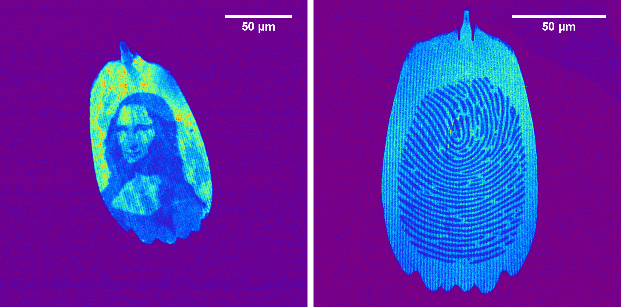
Arbitrary patterns inscribed in the butterfly wing scales by selective photo bleaching
|
Show more...
Concept of nonlinear laser scanning microscopy NLSM is an imaging technique, similar to confocal microscopy, where the laser beam is scanned across the specimen, the laser beam interact with the sample material and the signal is detected by a photodetector and recoreded on the computer. The image is formed on the computer from the recorded data. Unlike confocal microscopy where continuous lasers are used, in NLSM the ultrashort laser pulses are utilized. Due to high peak power of ultrashort laser pulses (~100 fs duration) the nonlinear optical effects become detectable. In the same time the average power of the laser beam is low enough to keep the samples undamaged.
|
Two Photon Excitation Fluroescence - TPEF is the most common effect/signal that is detected in NLSM. It is analogous to the single photon excitation that is utilized in confocal and epifluroescent microscopy, but in TPEF the fluorescent molecule is excited by absorbing two photons simultaneously. Upon the two photon excitation, the fluorescence occurs in the same manner as in the single photon excitation.
|
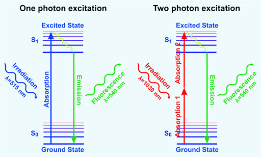
Principle of two photon excitation
|
In addition to TPEF, two more nonlinear optical effects can be detected in NLSM, that do not have analog in confocal microscopy. Those are Second Harmonic Generation - SHG and Third Harmonic Generation - THG.
Adventages and applications of NLSM The laser beam is usually focused by high numerical aperture (NA) objective in order to have as tight as possible focusing. Due to tight focusing and ultra short duration of the laser pulse, the signals occur only in very confined region, i.e. in focal volume, enabling imaging:
- with no (S/THG) or reduced (TPEF) photo bleaching, photo damage and photo toxicity
- at large penetration depths and high axial resolution
- without descanning and pinhole filtering
- label free imaging (SHG, THG and auto TPEF)
NLSM is indispensable technique for in vivo imaging!
Further readings on NLSM:
- NLSM, all modalities:
- TPEF:
- Two-photon excitation microscopy, text in Wikipedia
- Two-photon Fluorescence Light Microscopy, Peter TC So, Massachusetts Institute of Technology, Cambridge, Massachusetts, USA
- Principles of Two-Photon Excitation Microscopy and Its Applications to Neuroscience, Karel Svoboda and Ryohei Yasuda. Neuoron 50, 823 (2006)
- Two-photon excitation of fluorescence for three-dimensional optical imaging of biological structures, Alberto Diaspro and Mauro Robello.
Journal of Photochemistry and Photobiology B: Biology 55, 1 (2000)
- SHG:
- Second-harmonic imaging microscopy, text in Wikipedia
- Backward second harmonic generation from starch for in situ, real time pulse characterization in multiphoton microscopy, Anisha Thayil K. N et al., Proceedings of SPIE - The International Society for Optical Engineering 6442
- Membrane imaging by simultaneous second-harmonic generation and two-photon microscopy, Laurent Moreaux, Olivier Sandre, Mireille Blanchard-Desce, Jerome Mertz, Optics Letters 25 (5), 320 (2000)
- Second harmonic generation microscopy for quantitative analysis of collagen fibrillar structure, Xiyi Chen, Oleg Nadiarynkh, Sergey Plotnikov, Paul J Campagnola. Nature Protocols 7, 654 (2012)
- THG:
- Third harmonic generation microscopy, Jeff A. Squier, Michiel Müller, G. J. Brakenhoff, and Kent R. Wilson. Optics Express 3, 315 (1998)
- Imaging lipid bodies in cells and tissues using third-harmonic generation microscopy, Nature Methods 3(1):47-53 (2006)
- Third harmonic generation microscopy of cells and tissue organization, Bettina Weigelin, Gert-Jan Bakker, Peter Friedl. Journal of Cell Science 129, 245 (2016)
- Nonlinear optical phenomena:
- Ultrafast (femtosecond) lasers:

Lasers
Ti:Sa
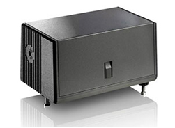
Pulse picker
Yb:KGW
GLX-Yb, Time-bandwidth producs AG - SESAM mode locked Yb KGW laser wavelength - 1040nm
- pulse duration - 200 fs
- repetition rate - 83 MHz
- maximal average power - 2 W

Optical autocorrelator:
Home-made optical autocorrelator for ultrashort pulse duration measurements. Built up through the
bilateral projects with DESY.
- Michelson interferometer with retroreflctor (corner cube prism) in the arm with motorized stage
- second order (intensity) autocorrelation using BBO crystal
- pulse durations: 10 fs - 100 ps
- computer controled
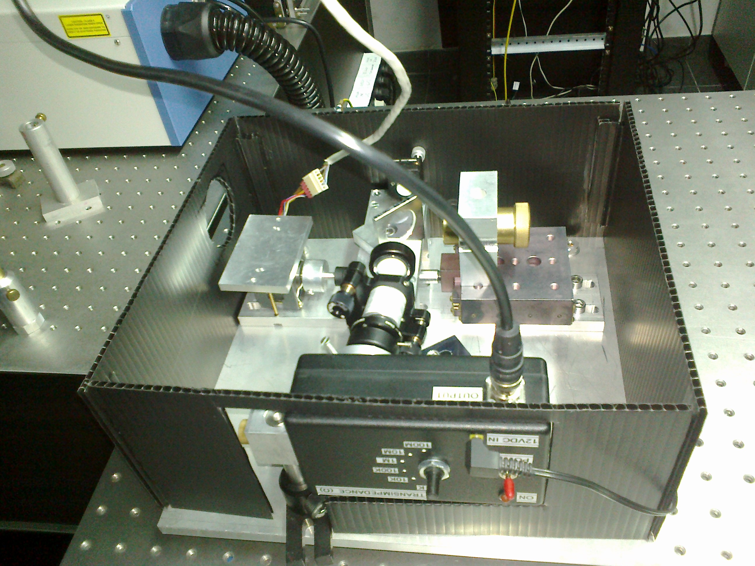
Optical autocorrelator set up
Control software inteface and autocorrelation trace for: (left) fs pulse from Mira 900; and (right) uncompressed ps pulse from RegA 9000

Cooled CCD spectrometer for weak signal measurements
Sensitive, fiber coupled USB spectrometer with high SNR suitable for the detection and analysis of weak fluorescence originating from the biological samples.
- Glacier X, bz BWTEK
https://bwtek.com/products/glacier-x/
- Coupled with NLSM setup
- Spectral analysis of the fluorescence signal upon the excitation in the arbitrary chosen point on the image
- Resolution: 4 nm
- Range: 350-150 nm
Purchased through the project
"Move For The Science" funded by
Filip Moriss.
